
What Do Dental X-Rays Show, and How Often Are They Needed?
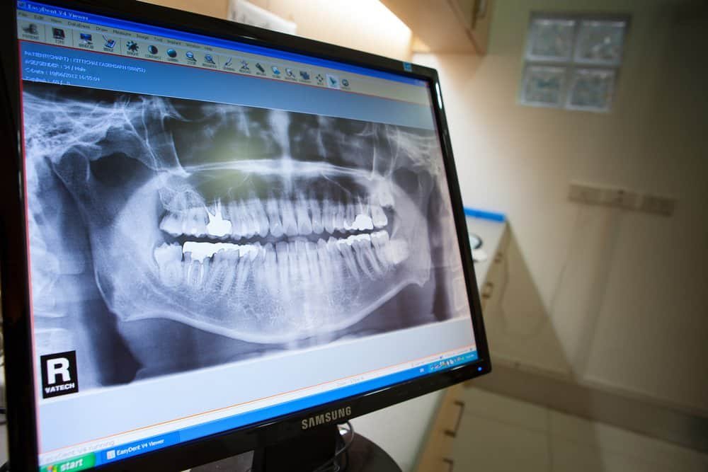
Dental X-rays can reveal cavities, roots, bone levels, impacted teeth and other details that are not visible during a clinical exam. How often they are needed depends on your symptoms, oral health, age, risk factors and prior images, not a fixed schedule for every patient.
The short answer
Dental X-rays are diagnostic tools. They help a dentist look between teeth, below the gumline and inside the jaws, where a visual exam cannot provide the whole picture. Different images answer different questions, so the right type and timing should be based on what your dentist needs to evaluate.
You may need an image sooner if you have pain, swelling, an injury, a suspicious area, extensive dental work, active gum disease or a higher risk of decay. If you are healthy and recent images still answer the clinical question, new X-rays may not be necessary at every visit.
The best dental X-ray is the one that answers a clinical question with the least necessary exposure.
What can dental X-rays reveal?
Dental X-rays can show changes that may be difficult or impossible to see directly. Depending on the image, they may help identify:
- Decay between teeth or beneath existing restorations.
- The roots of teeth and the surrounding bone, including changes near the root tip that may occur with infection.
- Bone levels around teeth, which can help evaluate periodontal disease.
- Impacted or developing teeth, including wisdom teeth.
- Jaw anatomy and the relationship of teeth to nearby structures.
- Problems after an injury or changes that need monitoring over time.
An X-ray is one part of diagnosis. It does not replace a clinical exam, and not every condition will appear clearly on an image. Your symptoms, medical and dental history, and what the dentist sees during the exam still matter.
Why are different types of dental X-rays used?
Each image has a different field of view and level of detail. A dentist chooses the type that is most likely to answer the question at hand.
Bitewing X-rays
Bitewings show the crowns of upper and lower back teeth together. They are commonly used to look for decay between teeth and to assess the height of the supporting bone.
Periapical X-rays
A periapical image shows an entire tooth from crown to root tip, plus nearby bone. It can help evaluate pain, infection, root anatomy, trauma and treatment around a specific tooth.
Panoramic X-rays
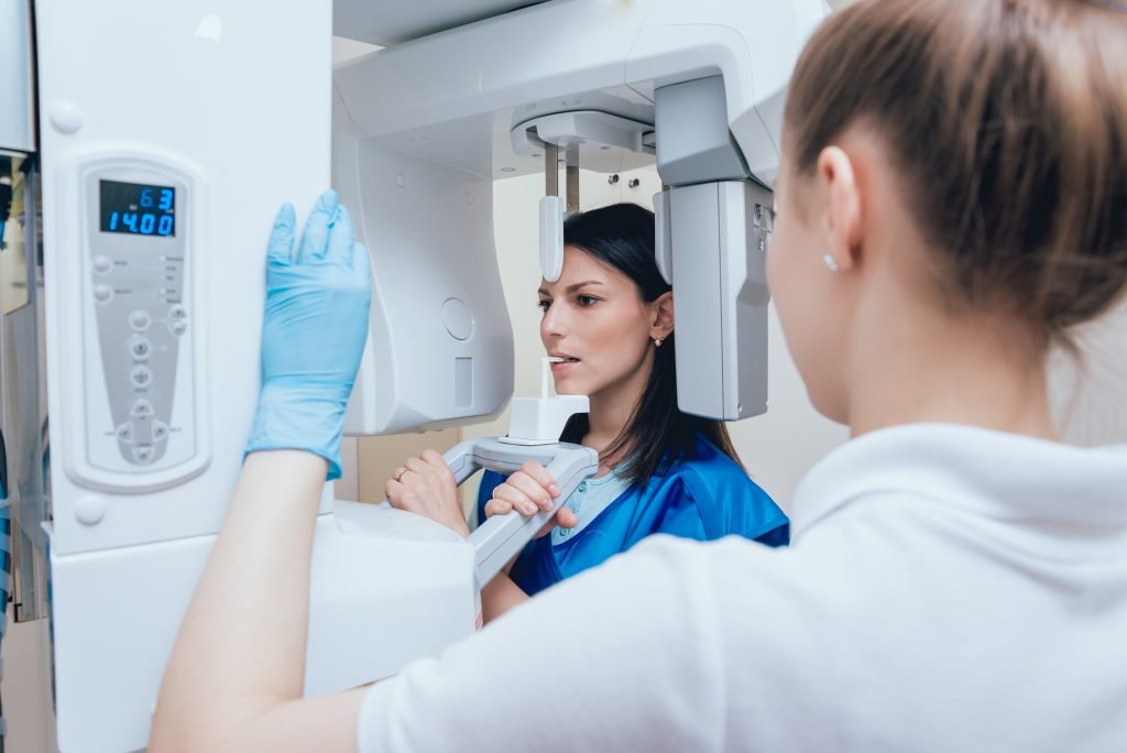
A panoramic X-ray captures a broad view of the teeth and jaws in one image. It can be useful for evaluating wisdom teeth, development, jaw relationships and other larger areas. Because it provides a wide overview rather than the same fine detail as every intraoral image, it does not automatically replace bitewings or periapicals.
Cone-beam CT scans
A CBCT scan creates three-dimensional views. It may be selected when 3D information could affect diagnosis or planning. Because it provides more information and generally more radiation than a single routine 2D image, current guidance supports using it selectively rather than as a default screening test.
How often do adults need dental X-rays?
There is no single interval that is right for every adult. The American Dental Association’s updated 2026 recommendations emphasize taking radiographs only when they are clinically necessary after reviewing the patient’s history, prior images and current exam.
Timing may depend on:
- whether you are a new patient and what recent records are available;
- new pain, swelling, trauma or other symptoms;
- your current risk of developing cavities;
- gum health and any history of bone loss;
- existing fillings, crowns, implants or root canal treatment that may need evaluation;
- age, growth and tooth development; and
- whether older images still show the area clearly enough to guide care.
A visit for a dental exam does not automatically mean that every type of X-ray is due. If you are returning after a long gap, bringing recent images from another office may help avoid unnecessary repeats.
Are dental X-rays safe?
Dental X-rays use ionizing radiation, so every image should be justified and optimized. Modern digital systems, careful positioning, the smallest useful field of view and appropriate exposure settings help keep doses low. The goal is to obtain the diagnostic information needed without taking images that are unlikely to change care.
The ADA’s 2024 safety recommendations also state that routine abdominal shielding and thyroid collars are generally no longer recommended when modern equipment and evidence-based techniques are used. Shielding can sometimes interfere with the image and lead to a repeat exposure. If you have questions or concerns, ask why an image is recommended, what area it will show and how the office limits exposure.
Tell the dental team if you are pregnant or may be pregnant. Necessary dental imaging can still be considered when it will affect care, but the decision should be individualized.
What should I tell the dentist before imaging?
Share any recent dental X-rays from another office if they are available. Also mention new pain, sensitivity, swelling, injury, changes in your medical history, pregnancy or the possibility of pregnancy. These details help the dentist decide whether an image is needed and which type is most useful.
If it has been years since your last visit, you can read what to expect when returning to the dentist after a long gap. The first step is still an exam and a conversation, not an automatic assumption about which images you need.
What affects the cost of dental X-rays?
Cost varies with the type and number of images, whether a 2D or 3D study is needed, insurance benefits and whether usable recent records can be transferred. A limited image of one tooth is different from a full-mouth series, panoramic image or CBCT scan. Before imaging, you can ask which study is recommended, why it is needed and what your expected share may be. Veranda Family Dentistry does not list a universal X-ray price because the appropriate study depends on the clinical situation.
Questions patients often ask
Do I need dental X-rays at every visit?
Not necessarily. The timing should be based on your symptoms, current exam, risk factors and whether recent images still provide useful information.
Can an X-ray find every cavity?
No single image or test finds every problem. Bitewings can be especially helpful for decay between back teeth, but a clinical exam and other information are still needed.
Can a panoramic image replace bitewings?
Not always. A panoramic image gives a broad overview, while bitewings are designed to show specific tooth surfaces and bone levels in greater detail. The two studies answer different questions.
Why would a dentist recommend a CBCT scan?
A CBCT may be recommended when three-dimensional information could affect diagnosis or treatment planning. It should be selected for a specific clinical reason, not used automatically for routine screening.
Have a question about a recommended X-ray?
If you have dental pain, swelling, an injury or questions about recent imaging, call Veranda Family Dentistry in Port Saint Lucie at 772.336.2300. We can help you arrange an evaluation and explain the purpose of any image recommended after your exam.
Sources and further reading
- American Dental Association: Updated recommendations for patient selection and dental radiography, January 5, 2026
- American Dental Association: Radiation safety and dental radiography recommendations
- U.S. Food and Drug Administration: Selection of patients for dental radiographic examinations
This article is general educational information and is not a diagnosis. Recommendations depend on an individual examination and health history.
Request an appointment
Use the form below to request a visit. For urgent symptoms, please call 772.336.2300.